45 the human heart and its labels
The Anatomy of the Heart, Its Structures, and Functions The heart is the organ that helps supply blood and oxygen to all parts of the body. It is divided by a partition (or septum) into two halves. The halves are, in turn, divided into four chambers. The heart is situated within the chest cavity and surrounded by a fluid-filled sac called the pericardium. This amazing muscle produces electrical ... Heart Anatomy: Labeled Diagram, Structures, Blood Flow ... - EZmed Image: Use the 2x2 table to label the 4 chambers of the heart, including the right atrium, right ventricle, left atrium, and left ventricle. Tricuspid Valve and Mitral Valve Now that we have a good understanding of the 4 chambers of the heart, let's move on to the 4 main valves.
Diagram of Human Heart and Blood Circulation in It Exterior of the Human Heart A heart diagram labeled will provide plenty of information about the structure of your heart, including the wall of your heart. The wall of the heart has three different layers, such as the Myocardium, the Epicardium, and the Endocardium. Here's more about these three layers. Epicardium

The human heart and its labels
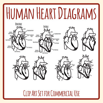
Human Heart Labeled Diagram The Human Heart Diagram Labeled - Human ... Jun 29, 2017 - Human Heart Labeled Diagram The Human Heart Diagram Labeled - Human Anatomy photo, Human Heart Labeled Diagram The Human Heart Diagram Labeled - Human Anatomy image, Human Heart Labeled Diagram The Human Heart Diagram Labeled - Human Anatomy gallery A Diagram of the Heart and Its Functioning Explained in Detail A well labeled human heart diagram given in this article will help you to understand its parts and functions. The human body is the best machine created by God. Every single part of our body is so well designed, that it works continuously throughout our life. CH. 20 Assessment Flashcards | Quizlet Identify the following structures of the heart. Label the components of the cardiac conduction system. Note that the human heart obtains a substantial amount of its energy from fats. The anterior side typically contains more adipocytes than the posterior side. Respond to the questions in the pop-up boxes addressing the structure of the wall of ...
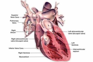
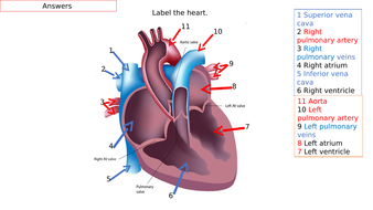
The human heart and its labels. Solved Label this frontal section of the human heart by - Chegg The arrows indicate direction of blood flow Right attium Aortic valve Lett pulmonary Pulmonary Superior vena Left ventricle artery valve Mitral valve Right pulmonary Pulmonary Tricuspid Inferior vena veis Right ventricle Loft atrium trunk valve cava This problem has been solved! See the answer Show transcribed image text Expert Answer How the Heart Works - The Heart | NHLBI, NIH The Heart. The heart is an organ about the size of your fist that pumps blood through your body. It is made up of multiple layers of tissue. Your heart is at the center of your circulatory system. This system is a network of blood vessels, such as arteries, veins, and capillaries, that carries blood to and from all areas of your body. A Labeled Diagram of the Human Heart You Really Need to See The human heart, comprises four chambers: right atrium, left atrium, right ventricle and left ventricle. The two upper chambers are called the left and the right atria, and the two lower chambers are known as the left and the right ventricles. The two atria and ventricles are separated from each other by a muscle wall called 'septum'. The Anatomy of the Heart - Quiz 1 - Free Anatomy Quiz The heart - an image of the heart with blank labels attached The circulatory system - upper body image, with blank labels attached The circulatory system - lower body image, with blank labels attached The circulatory system - a PDF file of the upper and lower body for printing out to use off-line Articles :
Human Heart - Diagram and Anatomy of the Heart - Innerbody The heart is a muscular organ about the size of a closed fist that functions as the body's circulatory pump. It takes in deoxygenated blood through the veins and delivers it to the lungs for oxygenation before pumping it into the various arteries (which provide oxygen and nutrients to body tissues by transporting the blood throughout the body). 13+ Heart Diagram Templates - Sample, Example, Format Download Human heart is a complicated figure and for students from science, they will often need the images of the heart for its illustration. The above collection of heart samples will make it easier for students to download, print and use it in their projects. The images with labels and detailed explanations can also be used in text books. Human Heart Diagram Labeled | Science Trends Human Heart Diagram Labeled Daniel Nelson 1, January 2019 | Last Updated: 3, March 2020 The human heart is an organ responsible for pumping blood through the body, moving the blood (which carries valuable oxygen) to all the tissues in the body. Without the heart, the tissues couldn't get the oxygen they need and would die. Heart: Anatomy and Function Heart. Your heart is the main organ of your cardiovascular system, a network of blood vessels that pumps blood throughout your body. It also works with other body systems to control your heart rate and blood pressure. Your family history, personal health history and lifestyle all affect how well your heart works. Appointments 800.659.7822.
Exam 5 Flashcards | Quizlet Thick, middle layer of the heart- Myocardium Covers surfaces of the heart valves- Endocardium Mostly composed of cardiac muscle cells- Myocardium The inner surface of the heart- Endocardium Also known as the visceral pericardium- Epicardium` Place the labels in their correct locations. Heart Labeling Quiz: How Much You Know About Heart Labeling? Here is a Heart labeling quiz for you. The human heart is a vital organ for every human. The more healthy your heart is, the longer the chances you have of surviving, so you better take care of it. Take the following quiz to know how much you know about your heart. Questions and Answers 1. What is #1? 2. What is #2? 3. What is #3? 4. What is #4? Heart Diagram with Labels and Detailed Explanation - BYJUS The human heart is the most crucial organ of the human body. It pumps blood from the heart to different parts of the body and back to the heart. The most common heart attack symptoms or warning signs are chest pain, breathlessness, nausea, sweating etc. The diagram of heart is beneficial for Class 10 and 12 and is frequently asked in the ... Easy way to draw human heart and label its main parts - YouTube Hai friends, In this video I am drawing a human heart in an eas way and labeling it's main parts .All the important parts are marked. ...
Given alongside is a diagram of the human heart showing its internal ... Given alongside is a diagram of the human heart showing its internal structure. Label the parts marked 1 to 6, and answer the following questions.Which chamb...
Human Heart - Anatomy, Functions and Facts about Heart The human heart is about the size of a human fist and is divided into four chambers, namely two ventricles and two atria. The ventricles are the chambers that pump blood and atrium are the chambers that receive blood. Among which both right atrium and ventricle make up the "right heart," and the left atrium and ventricle make up the "left heart."
Carefully study the diagram of the human heart with labels I II III and ... 15. Carefully study the diagram of the humanheart with labels (I), (II), (III) and (IV). Selectthe option which gives correct identificationand its main function. Page 36 of 53. (a) (I) Pulmonary artery: Carry oxygenatedblood from heart to lungs(b) (II) Pulmonary veins: Carry oxygenatedblood from lungs to the heart(c) (III) Aorta: Carry ...
Diagram of the human heart Images, Stock Photos & Vectors - Shutterstock 14,495 diagram of the human heart stock photos, vectors, and illustrations are available royalty-free. See diagram of the human heart stock video clips Image type Orientation Sort by Popular Anatomy Healthcare and Medical Diseases, Viruses, and Disorders Icons and Graphics heart medicine organ human body circulatory system diagram Next of 145
Label+Heart+Diagram+Worksheet | Heart diagram, Heart for ... - Pinterest 4 Best Printable Heart Diagram To Label. See 4 Best Images of Printable Heart Diagram To Label. Inspiring Printable Heart Diagram to Label printable images. Label Heart Diagram Worksheet Human Heart Diagram Printable Heart Diagram Blank Heart Diagram Worksheet. Printablee.
File:Diagram of the human heart (no labels).svg - Wikimedia File:Diagram of the human heart (no labels).svg. Size of this PNG preview of this SVG file: 498 × 599 pixels. Other resolutions: 199 × 240 pixels | 399 × 480 pixels | 499 × 600 pixels | 639 × 768 pixels | 851 × 1,024 pixels | 1,703 × 2,048 pixels | 533 × 641 pixels.

SANDRA GARRETT RIOS SIQUEIRA OAB/PE 12636 = TRAFICANTE DE DINHEIRO FALSO. AMIGA DO PCC. : SANDRA ...
Human Heart (Anatomy): Diagram, Function, Chambers, Location in Body The heart is a muscular organ about the size of a fist, located just behind and slightly left of the breastbone. The heart pumps blood through the network of arteries and veins called the...
How to Draw a Human Heart: 11 Steps (with Pictures) - wikiHow Label the parts of the heart if you'd to reference it for anatomy. If you're trying to identify parts of the heart for a class you're taking, it's good practice to draw the heart yourself and label each segment. You can refer to your textbook in order to label the: Aorta; Superior vena cava; Inferior vena cava; Right and left atria
PDF Anatomy of Heart Labeled and Unlabeled Images aortic arch left pulmonary artery left pulmonary veins auricle of left atrium left atrium circumflex artery (in atrioventricular sulcus) coronary sinus left ventricle (c) posterior view of the external heart © 2019 pearson education, inc. ascending aorta superior vena cava right pulmonary artery right pulmonary veins right atrium inferior vena …
How to Draw the Internal Structure of the Heart - wikiHow Make sure to label the following: Superior Vena Cava Inferior Vena Cava Pulmonary Artery Pulmonary Veins Left Ventricle Right Ventricle Left Atrium Right Atrium Mitral Valves Aortic Valves Aorta Pulmonic Valve (Optional) Tricuspid Valve (Optional) 6 To finish, label "The Human Heart" above the sketch. Community Q&A Search Add New Question Question
Label the heart — Science Learning Hub Label the heart Add to collection In this interactive, you can label parts of the human heart. Drag and drop the text labels onto the boxes next to the diagram. Selecting or hovering over a box will highlight each area in the diagram. Pulmonary vein Right atrium Semilunar valve Left ventricle Vena cava Right ventricle Pulmonary artery Aorta

SANDRA GARRETT RIOS SIQUEIRA OAB/PE 12636 = TRAFICANTE DE DINHEIRO FALSO. AMIGA DO PCC. : SANDRA ...
CH. 20 Assessment Flashcards | Quizlet Identify the following structures of the heart. Label the components of the cardiac conduction system. Note that the human heart obtains a substantial amount of its energy from fats. The anterior side typically contains more adipocytes than the posterior side. Respond to the questions in the pop-up boxes addressing the structure of the wall of ...
A Diagram of the Heart and Its Functioning Explained in Detail A well labeled human heart diagram given in this article will help you to understand its parts and functions. The human body is the best machine created by God. Every single part of our body is so well designed, that it works continuously throughout our life.
Human Heart Labeled Diagram The Human Heart Diagram Labeled - Human ... Jun 29, 2017 - Human Heart Labeled Diagram The Human Heart Diagram Labeled - Human Anatomy photo, Human Heart Labeled Diagram The Human Heart Diagram Labeled - Human Anatomy image, Human Heart Labeled Diagram The Human Heart Diagram Labeled - Human Anatomy gallery


.png)


Post a Comment for "45 the human heart and its labels"