40 microscope with labels and functions
Microscope Parts, Function, & Labeled Diagram - slidingmotion Microscope parts labeled diagram gives us all the information about its parts and their position in the microscope. Microscope Parts Labeled Diagram The principle of the Microscope gives you an exact reason to use it. It works on the 3 principles. Magnification Resolving Power Numerical Aperture. Parts of Microscope Head Base Arm Eyepiece Lens Amazon.com: My First Lab Duo Scope Microscope - Young … KIDS MICROSCOPE KIT -- This kids microscope kit, in addition to the user manual and experiment guide, includes 5 blank slides, 1 concavity (well) slide, 4 prepared slides, 2 bottles of stain, 5 slide labels, 5 cover glasses, 50 sheets of lens paper, 1 plastic transfer pipette, 1 plain wooden applicator, 1 cotton-tipped applicator, 1 plastic forceps, 1 plastic test tube and 1 plastic …
Compound Microscope Parts, Functions, and Labeled Diagram Compound Microscope Parts, Functions, and Labeled Diagram Parts of a Compound Microscope Each part of the compound microscope serves its own unique function, with each being important to the function of the scope as a whole.

Microscope with labels and functions
Microscope Parts, Functions, and Labeling Flashcards | Quizlet Start studying Microscope Parts, Functions, and Labeling. Learn vocabulary, terms, and more with flashcards, games, and other study tools. en.wikipedia.org › wiki › Electron_microscopeElectron microscope - Wikipedia An electron microscope is a microscope that uses a beam of accelerated electrons as a source of illumination. As the wavelength of an electron can be up to 100,000 times shorter than that of visible light photons , electron microscopes have a higher resolving power than light microscopes and can reveal the structure of smaller objects. Parts of the Microscope with Labeling (also Free Printouts) Let us take a look at the different parts of microscopes and their respective functions. 1. Eyepiece it is the topmost part of the microscope. Through the eyepiece, you can visualize the object being studied. Its magnification capacity ranges between 10 and 15 times. 2. Body tube/Head It is the structure that connects the eyepiece to the lenses.
Microscope with labels and functions. Microscope With Labeled Parts And Functions Microscope With Labeled Parts And Functions Microscope With Labeled Parts and Functions Written by Editor in General Biology Microscope is a revolutionized scientific instrument which is used in research laboratories to examine the small objects that are not clearly visible and can't be seen by the naked eye. rsscience.com › stereo-microscopeParts of Stereo Microscope (Dissecting microscope) - Rs' Science Stereo microscopes (also called Dissecting microscope) are branched out from other light microscopes for the application of viewing "3D" objects. These include substantial specimens, such as insects, feathers, leaves, rocks, sand grains, gems, coins, and stamps, etc. Functionally, a stereo microscope is like a powerful magnifying glass. Microscope Quiz: How Much You Know About Microscope Parts And Functions? Holds the slide in place. Moves the stage up and down for focusing. 3. Moves the stage slightly to sharpen the image. Holds the high and low power objectives. It can be rotated to change the magnification. Supports the microscope. 4. Is used to regulates the amount of light on the specimen. › internet › portalCanon U.S.A., Inc. | EOS Utility EOS Utility is an application that brings together functions to communicate with the camera. These functions include downloading and displaying images, remote shooting, and camera control for each setting. For download instructions follow the steps below. Have your camera's Serial Number ready before you begin.
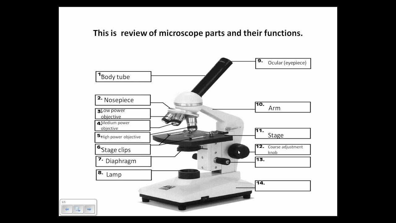
Microscope, Microscope Parts, Labeled Diagram, and Functions Illuminator: Illuminator is the most important microscope parts and it serve as light source for a microscope during slide specimen visualization. It is a continuous source of light (110 volts) used in place of a mirror. The mirror of microscope is used to reflect light from the external light source up through the bottom of the stage. Label Microscope Diagram - EnchantedLearning.com Using the terms listed below, label the microscope diagram. arm - this attaches the eyepiece and body tube to the base. base - this supports the microscope. body tube - the tube that supports the eyepiece. coarse focus adjustment - a knob that makes large adjustments to the focus. diaphragm - an adjustable opening under the stage, allowing ... Histology - Yale University It is white because the lipid is washed away during fixation and the vacuoles appear white under the microscope. Brown adipose tissue has smaller cells with many lipid droplets and mitochondria. It is brown because of the large number of cytochromes present. Distinguish the three kinds of cartilage. What type of collagen are they made of, and where are they found? … Label the microscope — Science Learning Hub 08.06.2018 · All microscopes share features in common. In this interactive, you can label the different parts of a microscope. Use this with the Microscope parts activity to help students identify and label the main parts of a microscope and then describe their functions.. Drag and drop the text labels onto the microscope diagram. If you want to redo an answer, click on the …
Parts of a Microscope Labeling Activity - Storyboard That Create a poster that labels the parts of a microscope and includes descriptions of what each part does. Click "Start Assignment". Use a landscape poster layout (large or small). Search for a diagram of a microscope. Using arrows and textables label each part of the microscope and describe its function. › 6-label-the-microscopeLabel the microscope — Science Learning Hub Jun 08, 2018 · All microscopes share features in common. In this interactive, you can label the different parts of a microscope. Use this with the Microscope parts activity to help students identify and label the main parts of a microscope and then describe their functions. Drag and drop the text labels onto the microscope diagram. If you want to redo an ... abberior-instruments.com › products › minfluxMINFLUX | Abberior Instruments By optimizing for low emission rates, the MINFLUX microscope zooms in on the molecule, concomitantly increasing the precision with which the molecular position is revealed. MINimizing fluorescence FLUXes by matching the dark center of the excitation beam with the molecule‘s position localizes molecules reliably and with 1-3 nanometer ... Microscope labeling and functions Flashcards | Quizlet Microscope labeling and functions STUDY Flashcards Learn Write Spell Test PLAY Match Gravity Created by mveet Terms in this set (27) Separates the eyepiece lens from the objective lenses Body Tube Holds the low-power and high-power objective lenses; allows the lenses to rotate for viewing Revolving Nosepiece Magnifies about 4x

Medical Pictures Info – Adrenal Gland
Microscope Parts & Functions - AmScope Microscope Parts and Functions Invented by a Dutch spectacle maker in the late 16th century, compound light microscopes use two sets of lenses to magnify images for study and observation. The first set of lenses are the oculars, or eyepieces, that the viewer looks into; the second set of lenses are the objectives, which are closest to the specimen.
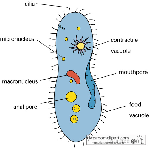
Science Clipart - paramecium_biology - Classroom Clipart
Labeling the Parts of the Microscope Labeling the Parts of the Microscope. This activity has been designed for use in homes and schools. Each microscope layout (both blank and the version with answers) are available as PDF downloads. You can view a more in-depth review of each part of the microscope here.
Microscope Review.wmv - YouTube
LAS X Industry Microscope software for Industry | Products Activate all relevant functions (e.g. for illumination settings, camera, measurements) with a few clicks ; Automatically store images on a regular basis with functionalities like autosave; Customizable user access. The software can handle multiple users who have different levels of microscope skills and diverse tasks to accomplish. Profiles according to user’s skills. The LAS …
Cell Structures as seen under the Light and Electron Microscope - Form ...
Compound Microscope: Definition, Diagram, Parts, Uses, Working ... - BYJUS Compound microscope is a type of optical microscope that is used for obtaining a high-resolution image. There are more than two lenses in a compound microscope. Learn about the working principle, parts and uses of a compound microscope along with a labeled diagram here.

Cell Types and Organelles
Microscope Parts and Functions - YouTube This video goes along with your microscope parts and function worksheet (Microscope Lab)
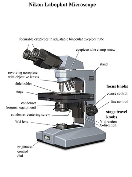
Using a Nikon Labophot compound microscope
Parts of Stereo Microscope (Dissecting microscope) – labeled … Compared to a compound microscope where the objectives attached to the nosepiece can be seen and identified individually (based on color bands and their respective labels), the objectives of a dissecting microscope are located in a cylindrical cone and, therefore, are not directly seen. For the stereo microscope that comes with multiple objective lens sets (fixed power style), the …

Anatomy and Physiology I Coursework: Microscope A+P
Confocal Microscopy - an overview | ScienceDirect Topics Shirley J. Wright, David J. Wright, in Methods in Cell Biology, 2002 B Disadvantages of Confocal Microscopy. Although confocal microscopes have many advantages, they do have some disadvantages. Confocal laser scanning microscopes (CLSMs) are limited by the available wavelengths of light produced by lasers (laser lines).This is unlike the conventional …
Vertebrate Histology Exam 2 Flashcards | Easy Notecards
22 Parts Of a Microscope With Their Function And Labeled Diagram A light microscope is a type of microscope that commonly uses visible light and a system of lenses to generate magnified images of small objects whereas electron microscope is a microscope that uses a beam of accelerated electrons as a source of illumination. It is a special type of microscope with a high resolution of images.

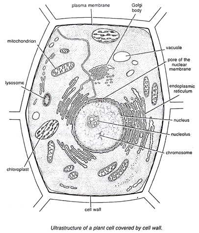
A LEVEL SCIENCE NOTES: Plant cell structure
Parts of a microscope with functions and labeled diagram Microscopes are instruments that are used in science laboratories to visualize very minute objects such as cells, and microorganisms, giving a contrasting image that is magnified. Microscopes are made up of lenses for magnification, each with its own magnification powers.
Post a Comment for "40 microscope with labels and functions"